Non-Surgical Treatment Options for Adenomyosis: What You Need to Know
The Telltale Symptoms of PAD/Leg Attack
PAD can manifest through a series of gradually intensifying symptoms. Initial stages of PAD often present as
- Intermittent claudication – pain, cramping or fatigue in the legs during physical activity, which subsides with rest. These sensations arise due to inadequate blood flow and oxygen delivery to the muscles. As PAD advances, individuals might experience pain even at rest, particularly in the calf, feet or toes, often disturbing sleep, and impairing quality of life.
- Non-healing ulcer- Beyond pain, other symptoms may also emerge. Diabetic patients may
present with non-healing wounds or ulcers on the feet or toes due to reduced blood flow,
leading to compromised tissue repair. - Coldness or numbness in the legs along with changes in skin colour (gangrene) are additional
indicators of restricted blood circulation. - Ignoring these signs can lead to further complications, including infections and in extreme cases, amputation.
How to diagnose PAD?
Detecting PAD requires a multifaceted approach, often involving medical history, physical examinations, and specialized tests. A key diagnostic tool is the Ankle-Brachial Index (ABI) test, which compares blood flow measurements in the arms and legs. A lower index suggests reduced blood flow to the legs, a hallmark of PAD. Imaging techniques like ultrasound, angiography, magnetic resonance angiography (MRA), and computed tomography angiography (CTA) provides visual insights into the extent of arterial narrowing and occlusion. Under an Interventional Radiologist, the patient can undergo diagnostic imaging and treatment.
Who are prone to develop PAD?
Older age, smoking, diabetes, high blood pressure, high levels of bad cholesterol, obesity, previous history of stroke or heart attack and a family history of cardiac diseases increase susceptibility to PAD. Many individuals dismiss early symptoms as a natural part of aging or attribute them to other causes.
Understanding Adenomyosis and How It Affects the Body
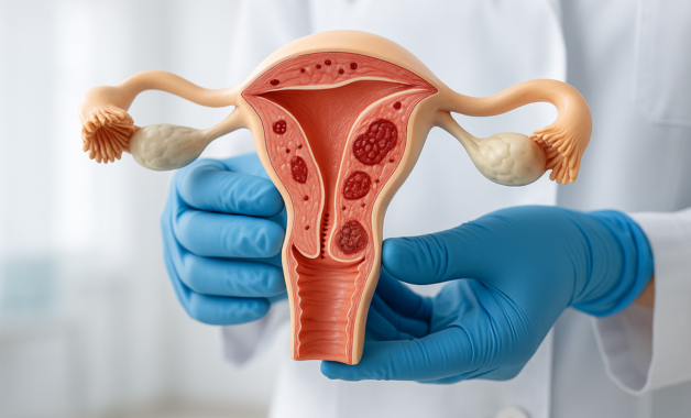
Adenomyosis occurs when the tissue that normally lines the uterus (endometrium) starts to grow into the muscular wall of the uterus (myometrium). This condition leads to an enlarged uterus and often results in severe menstrual cramps, excessive bleeding, pelvic pressure, and pain during intercourse. Unlike fibroids, which form as distinct growths, adenomyosis causes the uterus itself to become diffusely thickened, which can make it more difficult to detect and treat.
The condition most often affects women in their 30s and 40s, especially those who have had children. It can also be triggered or worsened by uterine trauma, such as that caused by surgeries like caesarean sections or dilation and curettage (D&C). Hormonal changes—particularly high levels of oestrogen—can fuel the growth of this displaced tissue, making the symptoms progressively worse with time.
The Limitations of Conventional Treatments
Many women begin their treatment journey with hormonal therapies such as birth control pills, progesterone-releasing intrauterine devices (IUDs), or gonadotropin-releasing hormone agonists (GnRH). While these can offer temporary symptom relief by controlling hormonal fluctuations, they often come with side effects and may not work in severe cases. In fact, once hormone therapy is stopped, symptoms often return.
When medication fails to offer sufficient relief, the conversation quickly shifts to surgical options. Hysterectomy, the surgical removal of the uterus, is frequently recommended as a definitive cure for adenomyosis. While this procedure does eliminate symptoms permanently, it is a major surgery with a long recovery period and permanent loss of fertility. For women who are not ready to undergo such a life-altering procedure, either due to personal reasons or the desire to preserve fertility, the search for an effective, non-surgical solution becomes essential.
Uterine Artery Embolization: A Breakthrough in Non-Surgical Management
In recent years, uterine artery embolization (UAE) has emerged as a highly effective, minimally invasive alternative for treating adenomyosis. This image-guided procedure is performed by an interventional radiologist and offers the distinct advantage of treating the condition without the need for general anaesthesia or open surgery.
The procedure involves inserting a thin, flexible tube through a small incision in the groin or wrist and guiding it into the uterine arteries using real-time imaging. Once in position, the radiologist injects tiny biocompatible particles that reduce blood flow to the affected areas of the uterus. With less blood supply, the adenomyosis-affected tissue gradually shrinks, easing symptoms like pain and heavy bleeding over time.
Uterine artery embolization is typically performed as a day-care or short-stay procedure. Most patients return home within 24 to 48 hours and can resume normal activities within a few days to a week. The uterus remains intact, which is a significant benefit for women who wish to retain their reproductive organs or avoid the emotional and physical impact of a hysterectomy.
Who Can Benefit from Adenomyosis Embolization Treatment?
This form of treatment is most suitable for women who have been diagnosed with adenomyosis—either focal or diffuse—through imaging techniques such as ultrasound or MRI. It is especially beneficial for those who experience debilitating symptoms but are not ready or willing to consider surgical options.
While uterine artery embolization is not traditionally recommended for women actively trying to conceive, several studies and case reports have documented successful pregnancies following the procedure. For women who are not currently planning to have children within 6 months of treatment, UAE presents a compelling alternative that offers lasting symptom relief with minimal disruption to daily life.
The success of this treatment depends on careful patient selection and accurate diagnosis. It is essential that women consult with both a gynaecologist and an interventional radiologist to fully understand their options and ensure that UAE is appropriate for their individual condition.
What Recovery Looks Like After Embolization
Recovery following uterine artery embolization is typically much quicker and easier than after traditional surgery. Some women may experience cramping, fatigue, mild fever, or nausea during the first few days—an expected reaction known as post-embolization syndrome. These symptoms are usually well-managed with oral medications and tend to resolve within a week.
Most women notice a reduction in menstrual bleeding within the first few cycles post-procedure, and many report a significant improvement in pelvic pain and pressure. In follow-up imaging, the uterus often returns to a more normal size, and the thickened adenomyotic areas begin to shrink. Regular monitoring is recommended over the following months to ensure that symptoms are improving and no complications arise.
A Look at Long-Term Outcomes and Effectiveness
Numerous studies have shown that uterine artery embolization offers lasting relief from the symptoms of adenomyosis in a majority of patients. According to published data, up to 75–85% of women experience significant reduction in pain and menstrual bleeding after the procedure. In some cases, additional treatments may be required years later, but the majority of patients avoid further intervention.
Unlike medication, which must be taken indefinitely, UAE offers a more definitive solution without removing the uterus. When performed by an experienced specialist, the risk of complications is low, and the overall success rate is high. Patients also appreciate the short recovery period, minimal discomfort, and the fact that they can return to work and regular activities much faster than after surgery.
Access to Treatment in Kerala and Beyond
In India, access to advanced interventional radiology procedures has significantly improved over the past decade. Women seeking non-surgical treatment for adenomyosis in Kerala can now receive high-quality care from experienced specialists using state-of-the-art technology.
At centres like GG Hospital and Sree Gokulam Superspeciality Hospital in Thiruvananthapuram, uterine artery embolization is performed under expert guidance, offering women a modern and safe solution close to home. Dr. Praveen Kesav Ramaswamy, a leading interventional radiologist in Kerala, has performed numerous embolization procedures, helping women reclaim their health and confidence without the need for major surgery.
Considering Your Options
Every woman’s experience with adenomyosis is different. While some may respond well to medication, others may find their symptoms worsening despite repeated attempts at conservative management. If you’ve been told that hysterectomy is your only option, it may be worth seeking a second opinion from a specialist in interventional radiology.
With the increasing availability of adenomyosis embolization treatment, women now have access to safer, more effective, and uterus-preserving options. Choosing the right treatment is a deeply personal decision, but knowing that surgery is not the only path forward can be incredibly empowering.
Consultation and Support
If you’re experiencing heavy periods, intense cramps, or pelvic pain and suspect adenomyosis, it’s important to undergo a proper diagnosis and discuss all available treatment options. Uterine artery embolization may be the alternative you’ve been searching for.
Dr. Praveen Kesav Ramaswamy offers advanced interventional radiology services for patients across Kerala. With specialised training from AIIMS and years of clinical experience, he is committed to delivering minimally invasive, life-changing treatments in a safe and supportive environment.
To learn more about non-surgical options for adenomyosis or to book a consultation, contact:
📞 Phone/WhatsApp: +91 9288023024
📧 Email: rpraveenkesav@gmail.com
📍 Locations: GG Hospital & Sree Gokulam Medical College, Thiruvananthapuram

")
